Microsporidia in fish caused by the species Microsporidia, which are a group of spore-forming unicellular parasites belonging to the phylum Microspora and the order Microsporidia.
These parasites have a wide host range, spanning protozoa and man, and are known to be responsible for many significant diseases.
In this blog article, we will delve into the causes, life cycle, symptoms, diagnosis, prevention, and treatment of Microsporidiosis in fish and crustaceans.
From their morphology to their geographical distribution and impact on various fish species, we will explore the different facets of this emerging disease.
What is Microsporidia
Microsporidia are a group of single-celled parasites that belong to the phylum Microsporidia. They are among the smallest known eukaryotic organisms, typically measuring between 1 and 5 micrometers in size.
Microsporidia are unique in that they lack certain organelles typically found in eukaryotic cells, such as mitochondria, and instead possess unique structures known as polar tubes that they use to invade host cells.
Microsporidia are intracellular parasites that can infect a wide range of animals, including humans, mammals, birds, fish, insects, and even other microorganisms.
Microsporidiosis is an emerging disease in hosts from aquatic and terrestrial biomes. Over 100 species of Microsporidia belonging to 21 genera have been identified as causing microsporidiosis in fish.
Transmission of Microsporidiosis
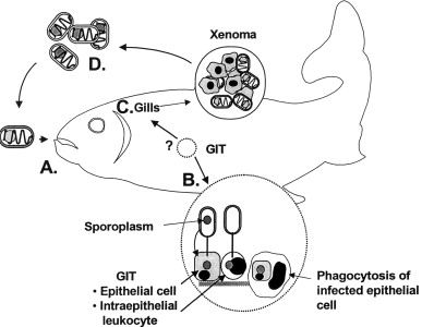
The transmission of infection to fish usually occurs through water, and common species causing microsporidiosis in fish include Pleistophora hyphessobryconis, Hetrosporis anguillarum, Kabatana sp., Microsporidium sp., and Perezia sp.
These spore-forming parasites affect the host tissues and form cysts that may be restricted by the connective tissue of the host.
Morphology of Microsporidia
The morphology of Microsporidia is unique and useful in distinguishing between different species.
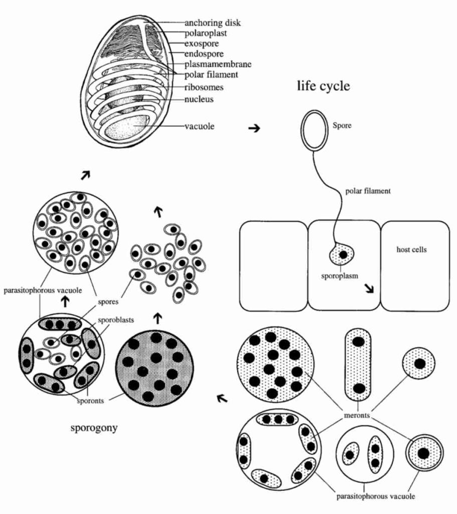
Some of the morphological features of microsporidia include the production of resistant spores that vary in size, possession of a unique organelle called the polar tubule or polar filament, and appearance of spores as ovoid and refractile on Gram stains, appearing bright purple.
The cuniculi cell wall and microsporidian spore wall contain glycosylated proteins, and microsporidia lack mitochondria but possess mitosomes.
They also lack motile structures such as flagella, and the spores are protected by a wall consisting of three layers with exospores having dense electone.
Life Cycle of Microsporidia

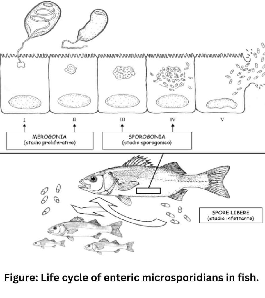
The life cycle of Microsporidia consists of two distinct stages, namely merogony or the proliferating stage, and sporogony or the infecting or mature stage.
Germination occurs, resulting in the extension of the polar tube and the injection of sporoplasm into the host cytoplasm by the polar tube, a specialized invasive structure.
The sporoplasm then develops into meronts, which is the proliferative stage where replication occurs through fusion by the formation of multinucleate plasmodium.
These meront cell membranes thicken and form sporonts, which develop into mature spores equipped with the apparatus.
The presence of the injection apparatus is a characteristic that defines microsporidia as a monophyletic taxon.
The host cells later become distended with mature spores, resulting in rupture and release of the spores to the outer milieu or nearby cells inside the body, thus allowing Microsporidia to multiply.

Geographical Distribution of Microsporidia
Microsporidia of fishes are widely distributed in different species and geographic locations.
While most fish microsporidia are host-specific, a few show broad host specificity. Cases of microsporidiosis have been reported in the Americas, Asia, Europe.
Symptoms
The microsporidia parasite can infiltrate the central nervous system and muscles of fish, potentially causing symptoms such as emaciation and skeletal deformities.
However, some fish may carry the parasite without showing any overt clinical signs, making it a sub-clinical infection.

Diagnosis of Microsporidiosis
Diagnosis of microsporidiosis in fish can be challenging as the symptoms may vary and can be similar to other diseases.
However, there are several methods that can be used for diagnosis, including:
Microscopic examination: Microsporidia spores can be observed under a microscope using special staining techniques. Spores are typically small and have distinct characteristics, such as a polar tube or filament, which can help in identifying the species.
Molecular techniques: Polymerase chain reaction (PCR) can be used to detect the presence of microsporidian DNA in fish tissues. This method is highly sensitive and specific, allowing for accurate identification of the species causing the infection.
Histopathology: Examination of fish tissues under a microscope can reveal characteristic changes caused by microsporidiosis, such as inflammation, necrosis, and cyst formation.
Immunological methods: Enzyme-linked immunosorbent assay (ELISA) and immunofluorescence assays can be used to detect specific antibodies against microsporidia in fish serum, indicating an active infection.
Prevention of Microsporidiosis
Preventing microsporidiosis in fish can be challenging, as the spores can be resistant to environmental conditions and can persist in water for extended periods.
However, there are some measures that can help in reducing the risk of infection:
Quarantine: Quarantining new fish before introducing them to an existing fish population can help in preventing the spread of microsporidia and other diseases.
Water management: Maintaining clean and well-filtered water can help in reducing the load of microsporidia spores in the environment.
Biosecurity measures: Implementing strict biosecurity measures, such as disinfection of equipment and proper disposal of fish mortalities, can help in preventing the introduction and spread of microsporidia.
Avoiding contaminated water sources: Avoiding the use of contaminated water sources, such as stagnant water or water from unknown or potentially contaminated sources, can help in preventing microsporidiosis in fish.
Treatment of Microsporidiosis
There are currently no specific treatments for microsporidiosis in fish. However, some general measures can be taken to support fish health and reduce the impact of the infection:
Quarantine and removal of infected fish: Isolating and removing infected fish from the population can help in preventing the spread of microsporidia to other fish.
Supportive care: Providing optimal water quality, nutrition, and stress reduction measures can help in supporting fish immune function and overall health, which may aid in their ability to fight off the infection.
Disinfection: Thoroughly disinfecting tanks, equipment, and water sources can help in reducing the load of microsporidia spores in the environment.
Microsporidia infection in humans
Microsporidia infection in humans, also known as microsporidiosis, is a parasitic infection caused by microscopic organisms called microsporidia. These parasites are part of the phylum Microsporidia, and they are known to infect a wide range of animals, including humans.
Microsporidia can enter the human body through various routes, such as ingestion of contaminated food or water, inhalation of spores, or direct contact with infected individuals or surfaces.
Once inside the human body, microsporidia invade host cells and replicate, causing damage to tissues and organs. Microsporidiosis most commonly affects the gastrointestinal tract, but it can also affect other organs, such as the eyes, respiratory tract, urinary tract, and muscles, depending on the species of microsporidia involved.
Symptoms of microsporidiosis in humans can vary depending on the location of the infection and the species of microsporidia involved. Common symptoms may include diarrhea, abdominal pain, nausea, vomiting, weight loss, fatigue, eye redness, respiratory symptoms, and skin lesions.
However, in some cases, microsporidiosis can be asymptomatic, especially in individuals with a healthy immune system.
Diagnosis of microsporidiosis in humans typically involves microscopic examination of clinical samples, such as stool, urine, or biopsy specimens, to identify the characteristic spores of microsporidia.
Molecular methods, such as polymerase chain reaction (PCR), may also be used for species identification. Treatment options for microsporidiosis in humans generally involve antiparasitic drugs, such as albendazole, fumagillin, or nitazoxanide, although the choice of medication may depend on the species of microsporidia involved and the severity of the infection.
In immunocompromised individuals, such as those with HIV/AIDS, management of the underlying immune deficiency may also be important.
Prevention of microsporidiosis in humans includes practicing good hygiene, such as washing hands thoroughly with soap and water, avoiding consumption of contaminated food or water, and taking precautions in healthcare settings to prevent transmission.
It is especially important for individuals with weakened immune systems, such as those with HIV/AIDS, to take extra precautions to minimize the risk of microsporidia infection.
FAQs
Can microsporidiosis be transmitted to humans?
Microsporidiosis is primarily a disease of animals, and the risk of transmission to humans is low.
Is microsporidiosis a common disease in fish?
Microsporidiosis is not a common disease in fish, but it can occur in various species of fish, both in wild and captive populations.
The prevalence and severity of microsporidiosis outbreaks in fish can vary depending on factors such as water quality, stress levels, and overall fish health management practices.
Can microsporidiosis be spread through contaminated water?
Yes, microsporidiosis can be spread through contaminated water. Microsporidia spores are resistant to environmental conditions and can persist in water for extended periods.
Using contaminated water sources, such as stagnant water or water from unknown or potentially contaminated sources, can increase the risk of microsporidiosis in fish.
References
- Franzen, C., & Müller, A. (1999). Molecular techniques for detection, species differentiation, and phylogenetic analysis of microsporidia. Clinical microbiology reviews, 12(2), 243-285.
- Rodriguez-Tovar, L. E., Speare, D. J., & Markham, R. F. (2011). Fish microsporidia: immune response, immunomodulation and vaccination. Fish & Shellfish Immunology, 30(4-5), 999-1006.
- Ryan, J. A., & Kohler, S. L. (2016). Distribution, prevalence, and pathology of a microsporidian infecting freshwater sculpins. Diseases of aquatic organisms, 118(3), 195–206. https://doi.org/10.3354/dao02974
- Yurakhno, V. M., Voronin, V. N., Sokolov, S. G., Malysh, J. M., Kalmykov, A. P., & Tokarev, Y. S. (2021). Genetic diversity of Loma acerinae (Microsporidia) from different fish hosts and localities – Short communication. Acta veterinaria Hungarica, 69(1), 38–42. https://doi.org/10.1556/004.2021.00012
- Schuster, C. J., Sanders, J. L., Couch, C., & Kent, M. L. (2022). Recent Advances with Fish Microsporidia. Experientia supplementum (2012), 114, 285–317. https://doi.org/10.1007/978-3-030-93306-7_1
- Weiss L. M. (2001). Microsporidia: emerging pathogenic protists. Acta tropica, 78(2), 89–102. https://doi.org/10.1016/s0001-706x(00)00178-9
- Didier, E. S., & Weiss, L. M. (2006). Microsporidiosis: current status. Current opinion in infectious diseases, 19(5), 485–492. https://doi.org/10.1097/01.qco.0000244055.46382.23
- Han, B., & Weiss, L. M. (2017). Microsporidia: Obligate Intracellular Pathogens Within the Fungal Kingdom. Microbiology spectrum, 5(2), 10.1128/microbiolspec.FUNK-0018-2016. https://doi.org/10.1128/microbiolspec.FUNK-0018-2016
- Ozkoc, S., Bayram Delibas, S., & Akısu, C. (2016). Evaluation of pulmonary microsporidiosis in iatrogenically immunosuppressed patients. İyatrojenik immün baskılanmış hastalarda pulmoner mikrosporidiosisin değerlendirilmesi. Tuberkuloz ve toraks, 64(1), 9–16. https://doi.org/10.5578/tt.10207
- Sanders, J. L., Watral, V., & Kent, M. L. (2012). Microsporidiosis in zebrafish research facilities. ILAR journal, 53(2), 106–113. https://doi.org/10.1093/ilar.53.2.106
- Gross U. (2003). Treatment of microsporidiosis including albendazole. Parasitology research, 90 Supp 1, S14–S18. https://doi.org/10.1007/s00436-002-0753-x
- Phelps, N. B., Mor, S. K., Armién, A. G., Pelican, K. M., & Goyal, S. M. (2015). Description of the microsporidian parasite, Heterosporis sutherlandae n. sp., infecting fish in the Great Lakes Region, USA. PLoS One, 10(8), e0132027.