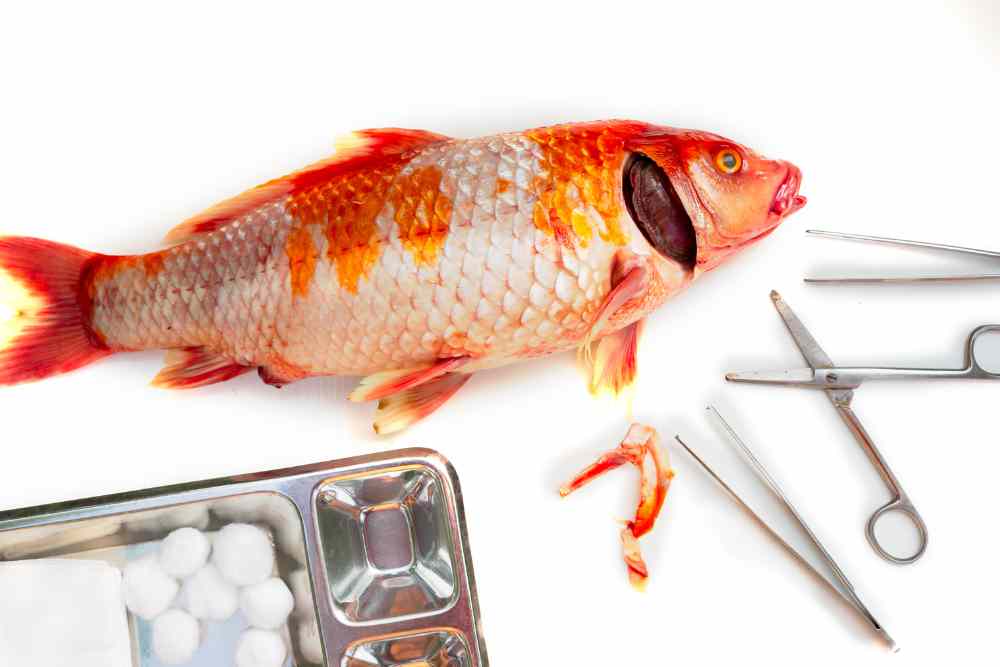
Fish disease diagnosis can is a complex and intimidating process. However, with the right information, knowledge, and resources, anyone can learn to diagnose fish diseases with confidence.
This article provides a beginner’s overview of fish disease diagnosis, offering vital tips and advice to increase your knowledge in this area.
You will learn key identification techniques, how to understand the signs of disease, how to identify disease causing agents and what actions to take if you identify a problem.

What is Diagnosis?
The word diagnosis derived from the Greek “dia” means through and “gnosis” means knowledge. Diagnosis the determination of the nature of the cause of a disease.
Disease diagnosis is the process of identifying a disease or medical condition in an individual based on its signs, symptoms, and medical history.
The diagnostic process may include a physical examination, laboratory tests, imaging studies, or other specialized procedures, depending on the suspected disease or condition.
The goal of disease diagnosis is to accurately identify the underlying cause of a patient’s symptoms and provide appropriate treatment to alleviate or manage the condition.
Objectives of Fish Diseases Diagnosis
The objectives of fish disease diagnosis are:
Identification of the causative agent: The primary objective of fish disease diagnosis is to identify the causative agent of the disease or infection, such as bacteria, viruses, parasites, or fungi.
Confirmation of the disease: Once the causative agent has been identified, the next objective is to confirm the presence of the disease or infection.
Determination of the severity of the disease: The severity of the disease or infection can be determined through laboratory tests, clinical signs, and symptoms.
Development of an appropriate treatment plan: Based on the identified causative agent and the severity of the disease, an appropriate treatment plan can be developed to manage the disease and minimize its spread.
Prevention of disease spread: Farmed fish are also at risk from disease, with the potential for mass die-offs if a disease outbreak is not quickly controlled.
Fish disease diagnosis can also help to prevent the spread of the disease or infection to other fish populations or aquatic environments.
Protect wild fishes: Fish diseases can cause major problems for both wild and farmed fish populations.
In wild populations, diseases can lead to a decline in numbers, as well as genetic diversity. This can have a knock-on effect on the whole ecosystem.
Protection of public health: Some fish diseases can be transmitted to humans, so fish disease diagnosis can also play a role in protecting public health.
Minimize economic loss: In addition to the economic impacts, fish diseases can also have negative environmental impacts. Some diseases can cause mass die-offs of fish, which can lead to pollution and degraded water quality.
Fish diseases can have a devastating effect on aquaculture, causing significant financial losses. Early diagnosis is essential for effective treatment and prevention of the spread of disease.
Diagnostic Methods of Fish Diseases
Fish disease diagnosis broadly divided into two categories;
- Clinical or Presumptive Diagnosis
- Laboratory or Confirmatory Diagnosis
Clinical or Presumptive Diagnosis of Fish Disease
There are many different ways to diagnose fish diseases. One of the most important things to do when diagnosing fish diseases is to examine the fish carefully for any signs of illness.
Clinical diagnosis followed the two changes in fish;
- Changes in Behaviors
- Changes in Appearance
1. Changes in Behavior of Sick Fish
These changes in behavior can be due to the fish’s illness itself, or as a result of the stress of being ill.
Here is a table depict the different behavioral changes observed in diseased fish compared to healthy fish.
| Observation | Healthy fish | Diseased fish | Possible causes |
| Swimming | Normal | Erratic | Virus, Bacteria, Arugulas sp., Toxic substances |
| Swim freely and evenly | Swimming near the dike and exposing upside down | Swim bladder issue, gas bubble issue | |
| Normal | Unusual whirling swimming | Virus, bacteria, protozoa, nutritional imbalance | |
| Movement | Normal | Scrapping body against bottom, side of container, or hard object | External parasite (protozoa, Gyrodactylus etc.), crustacean parasite (Lernaea, Argulus etc.) |
| Move in every part of his territory | Fish congregate near water inlet, gasping | Low dissolved oxygen, trauma | |
| Active, vibrant | Fish become restless | Bacteria, protozoa | |
| Active, alert and sociable | Lying bottom of the tank | Gas bladder issue, parasite | |
| Swim different column of water | Unable to swim properly, sunken | Swim or air bladder disorder | |
| Feeding | Quick and enthusiastic | Reduced feeding | Bacteria, virus, parasite |
| Normal | Stop feeding | Virus, bacteria, stress | |
| Weight | Normal | Reduced | Bacteria, virus, parasite, stress |
| Normal | Lean body bigger head | Parasite, nutritional deficiency |
2. Changes in Appearance of Sick or Stressed Fish
| Observation | Healthy Fish | Diseased Fish | Possible causative agents & causes |
| Eye | Parallel with body shape | Bulging eye (pop eye\exophthalmia) | Bacteria, parasite, trauma |
| Bright and clear | Cloudy or milky eye (corneal opacity) | Bacteria, virus, parasite, trauma | |
| Gill Shape | Organized gill lamellae | Gill lamellae clubbed and irregular | Bacteria, parasite, trauma |
| Normal | Hemorrhage, Inflammatory gill | Bacteria, parasite, trauma | |
| Gill Color | Reddish or pink | Black & slimy | Bacteria, fungus |
| Normal bright color | Pale pink or white or mottled | Virus, bacteria, gill fluke, anemia | |
| Normal red or pinkish red | Bright red or hemorrhage | Excess ammonia | |
| Normal red or pinkish red | Bright red | Bacteria, fungus, parasite | |
| Not slimy or dry and smell like fresh seaweed | Excess slime and rotten flavor | Bacteria, degraded water quality | |
| Rhythmically open and close gill cover | Gill cover opened continuously | Parasite, trauma | |
| Body Color | Bright color | Posterior dark coloration | Lead poisonings |
| Bright color | Pale or faded body color | Bacteria, virus, nutritional deficiency | |
| Bright color | Hemorrhagic patches | Bacteria, virus | |
| Natural color & spot | Tiny white spot on body | Ich parasite, fungus | |
| Scale | Parallel with body skin | Protruded and spiky | Bacteria, stress |
| Fin & Tail | Not spiky | Fins with spiky naked fin rays | Bacteria, protozoa, rubbing |
| Normal | |||
| Normal | Fins eaten away, edges leveled off | Cannibalism, bacteria, fungus | |
| Abdomen | Parallel with body shape | Bloated or distended abdomen (Ascites), dropsy | Bacteria, virus, helminth parasite, pregnant, over feeding |
| Normal | Sunken | Nutritional deficiency | |
| Anus | Clear vent, no stringy faces | Stringy faces | Nutritional disorder, intestinal parasite |
Laboratory Diagnosis of Fish Diseases
Laboratory diagnosis can be conducted through any of the or combination of the following process,
- Microscopic observation
- Histopathological diagnosis
- Microbiological diagnosis
- Immunological\Serological diagnosis
- Molecular diagnosis
1. Microscopic Observation
Microorganisms can be diagnosed through a microscope by observing their physical characteristics such as size, shape, and staining properties.
Microscopes allow us to view microorganisms at a high magnification, which makes it easier to identify and classify them based on their morphological features.
One of the primary methods of identifying microorganisms is through Gram staining, which involves staining the cell wall of bacteria with crystal violet and iodine, followed by a counterstain of safranin.
This staining process allows for the differentiation of bacteria into two main groups: Gram-positive and Gram-negative. Gram-positive bacteria appear purple under the microscope, while Gram-negative bacteria appear pink.
Other staining techniques, such as acid-fast staining, are used to identify specific groups of bacteria, such as mycobacteria, which have a unique cell wall structure that makes them resistant to standard staining methods.
In addition to staining techniques, microscopy allows for the observation of various structural features of microorganisms, such as flagella, pili, and spores, which can aid in their identification.
Overall, microscopy is a powerful tool for diagnosing microorganisms and provides valuable information about their morphology and characteristics, which can be used to guide treatment and control strategies.
2. Histopathological Diagnosis
Histopathological diagnosis is the process of examining and analyzing tissue samples under a microscope to diagnose disease.
It involves the study of the structural and cellular changes that occur in tissues due to disease, injury, or other pathological conditions.
In histopathological diagnosis, a small piece of tissue, called a biopsy, is taken from the patient and then processed and prepared for examination under a microscope.
The tissue sample is usually stained to enhance the contrast and visibility of different structures and cells within the tissue.
A pathologist then examines the tissue sample under a microscope and looks for characteristic features of the disease.
This may involve the identification of abnormal cells, changes in tissue structure, or the presence of inflammation, infection, or other pathological changes.
Histopathological diagnosis is used in the diagnosis of a wide range of diseases, including cancer, infections, autoimmune diseases, and genetic disorders.
It is a critical tool in the diagnosis, prognosis, and treatment of many diseases, as it can provide important information about the nature and severity of the disease, as well as guide treatment decisions.
Microbiological Diagnosis
Microbiological diagnosis is an important aspect of fish disease diagnosis, which involves the identification of the specific microorganisms responsible for causing the disease in fish.
Microbial culture is a fundamental technique in microbiology that allows scientists to study the growth and behavior of bacteria in a controlled laboratory setting.
In this technique different culture media and environmental conditions are used to identify the specific bacteria, viruses, fungi, or parasites responsible for causing the disease.
Once the microorganism is identified, it can be further characterized through various tests, such as antibiotic susceptibility testing, to determine the best treatment option.
Molecular Diagnosis of Fish Diseases
Molecular tests are powerful tools that can be used to identify pathogenic bacteria in the laboratory by analyzing their DNA or RNA.
These tests can provide rapid and accurate identification of bacterial species, which can be critical for effective treatment of bacterial infections.
Polymerase Chain Reaction (PCR)
PCR is a technique that amplifies specific regions of bacterial DNA, allowing for the detection and identification of the bacterial species.
This method is rapid and highly sensitive, allowing for the detection of even small amounts of bacterial DNA.
PCR can be used to amplify and detect specific DNA sequences that are unique to the microorganism causing the disease, even in very small quantities.
DNA Sequencing
DNA sequencing can be used to determine the sequence of the bacterial DNA, which can provide information on the bacterial species and any mutations that may be present.
Whole-Genome Sequencing (WGS)
Whole-genome sequencing analyzes the entire bacterial genome, providing detailed information on the bacterial species and any genetic variations that may be present.
DNA Barcoding
DNA barcoding is a molecular technique that can be used to identify pathogenic microbes in fish.
This technique involves amplifying a specific region of the microbe’s DNA, which is then sequenced and compared to a reference database to identify the microbe.
In the case of fish disease diagnosis, DNA barcoding can be used to identify the specific microbe responsible for causing the disease.
This is particularly useful when traditional methods, such as microbial culture or biochemical tests, are unable to identify the microbe.
The first step in DNA barcoding is to select a suitable DNA marker that can be used to identify the microbe.
For pathogenic microbes, a suitable marker might be a conserved region of the genome that is unique to the species or strain causing the disease.
Once a suitable marker has been selected, DNA is extracted from the sample (e.g. fish tissue or swab) and the marker is amplified using PCR.
The resulting DNA fragment is then sequenced and compared to a reference database, such as GenBank or the Barcode of Life Data Systems (BOLD).
The reference database contains DNA sequences from known pathogenic microbes, allowing for the identification of the microbe causing the disease in the fish.
This information can then be used to develop appropriate treatment and management strategies to control the disease.
DNA Microarray
The DNA microarray technique is a powerful tool that can be used to identify pathogenic microorganisms in fish.
This technique involves the use of a small glass slide or chip that contains thousands of different DNA probes that are specific to different microorganisms.
To identify the pathogenic microorganism, a sample of the fish tissue or fluid is collected and the DNA is extracted. The extracted DNA is then labeled with a fluorescent tag and added to the DNA microarray chip.
The labeled DNA hybridizes with the specific DNA probes on the chip, producing a fluorescent signal that can be detected and analyzed.
By comparing the pattern of fluorescence to a reference database of known microorganisms, the pathogenic microorganism can be identified.
This database can include a wide range of microorganisms, including bacteria, viruses, fungi, and parasites.
One of the key advantages of the DNA microarray technique is that it can detect multiple microorganisms simultaneously, making it a powerful tool for the rapid diagnosis of fish diseases caused by multiple pathogens.
It is also a high-throughput technique, which means that it can process a large number of samples in a relatively short period of time.
In addition to its diagnostic applications, DNA microarray technology can also be used to monitor the presence and abundance of microorganisms in fish farms or aquatic environments, providing important information for disease management and control.
Serological or Immunological Diagnosis of Fish Diseases
Serological or Immunological tests can be used to identify pathogenic bacteria by detecting the presence of bacterial antigens or antibodies in the body.
These tests rely on the specific binding of antibodies to bacterial antigens, allowing for the identification and quantification of the bacteria.
It involve the use of antibodies, which are proteins that can bind specifically to a particular antigen, such as a bacterial cell surface protein.
Here are some common immunological tests used to identify pathogenic bacteria:
Enzyme-linked immunosorbent assay (ELISA)
This test uses a specific antibody that is linked to an enzyme.
This test can be used to detect specific bacterial species or to quantify the immune response to an infection.
When the antibody binds to the antigen on the surface of the pathogenic bacteria, the enzyme is activated and can be detected using a colorimetric or fluorescent signal.
Western blotting
Western blotting is a technique that separates bacterial proteins by size and then detects specific proteins using antibodies.
This technique can be used to identify bacterial species and to detect specific virulence factors.
Immunofluorescence: Immunofluorescence uses fluorescent-labeled antibodies to detect the presence of bacterial antigens or antibodies in a sample.
This technique can be used to visualize the location of the bacteria in tissue samples or to detect the presence of specific bacterial species.
Agglutination Test
This test involves mixing bacterial cells with specific antibodies, which cause the cells to clump together or agglutinate.
The agglutination reaction can be observed visually and is used to identify specific bacterial species.
Rapid diagnostic tests
Rapid diagnostic tests use immunological techniques to detect specific bacterial antigens in patient samples.
These tests are often used for point-of-care diagnosis of bacterial infections, such as strep throat or urinary tract infections.