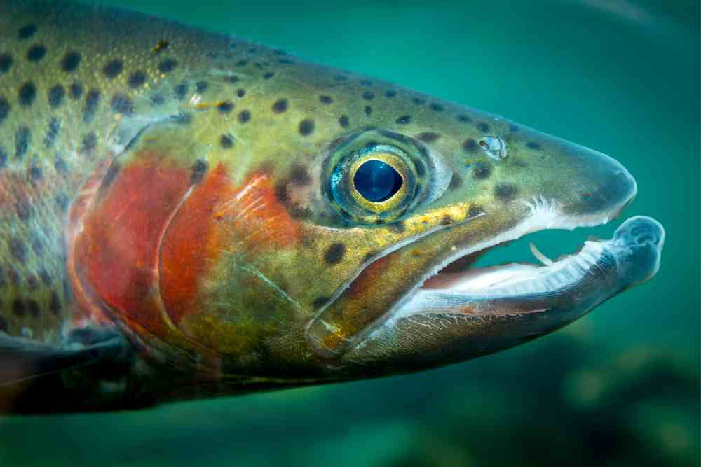
Enteric Redmouth Disease (ERM) is a serious bacterial infection of fish that can cause major health problems for freshwater and saltwater fish species.
It is caused by a bacterium named Yersinia ruckeri, and can have devastating effects on different species of fish, including trout and salmon and also called Yersiniosis.
It affects a variety of both wild and farmed fish species in different parts of the world, from North America to Europe and Asia.
It has a wide host range, broad geographical distribution, and causes significant economic losses in the fish aquaculture industry.
This article provides an overview of Enteric Redmouth Disease, including its causes, symptoms, and treatment options for those affected by it.

What Causes Red Mouth Disease?
Enteric Red Mouth Disease (ERM) is caused by the bacterium Yersinia ruckeri.
This bacterium is commonly found in fresh and brackish water environments and can infect a wide range of fish species, particularly salmonids such as rainbow trout, Atlantic salmon, and brook trout.
Morphologically, the ERM bacterium is a 1.0 x 2.0 to 3.0 um, gram negative, monotrichously flagellated rod which forms a smooth, circular, raised, entire, nonfluorescent, nonpigmented colony on nutrient agar with a buterous type of growth.
The bacterium enters the fish’s body through the mouth or gills and then multiplies rapidly in the intestine, causing inflammation and tissue damage.
This leads to the characteristic red discoloration of the mouth and fins, as well as other symptoms such as lethargy and loss of appetite.
ERM can be transmitted through several means, including contaminated water, equipment, or fish stock, and poor hygiene practices.
Stressors such as poor water quality, overcrowding, and suboptimal nutrition can weaken the fish’s immune system, making them more susceptible to infection.
ERM can affect fish from all age classes, but it is most acute in young fish (fry and fingerlings). The disease appears as a more chronic condition in older/larger fish.
Susceptible Species
Enteric red mouth disease, caused by the bacterium Yersinia ruckeri, can affect a wide range of fish species, but it is most commonly seen in salmonids, including:
- Rainbow trout (Oncorhynchus mykiss)
- Brown trout (Salmo trutta)
- Atlantic salmon (Salmo salar)
- Chinook salmon (Oncorhynchus tshawytscha)
- Coho salmon (Oncorhynchus kisutch)
- Steelhead trout (Oncorhynchus mykiss)
- Brook trout (Salvelinus fontinalis)
- Lake trout (Salvelinus namaycush)
- Cutthroat trout (Oncorhynchus clarkii)
- Arctic char (Salvelinus alpinus)
Other fish species that have been known to be affected by Enteric red mouth disease include pike, eel, perch, and whitefish.
Geographical Distribution of Enteric Redmouth Disease
Enteric red mouth disease has been observed in fish species in several countries worldwide.
Some of the countries where the disease has been reported include:
- Norway
- Canada
- United States
- United Kingdom
- Spain
- Germany
- France
- Sweden
- Finland
- Denmark
- Chile
- Japan
- Russia
- China
These are just a few examples of the countries where Enteric red mouth disease has been observed. It is important to note that the disease may be present in other countries or regions as well.
Enteric Red mouth Disease Symptoms
-The symptoms of enteric redmouth disease in fish include changes in behavior, such as swimming near the surface, lethargy, and loss of appetite.
-Darkening of the skin, subcutaneous hemorrhages around the mouth and throat, and exophthalmia.
-Infected fish may darken in color and have petechial hemorrhage of the visceral organs and tissues, including the liver, pancreas, adipose tissues, swim bladder, various coelomic mesenteries, and body musculature.
-Gross tissue edema may be present in the kidney, liver, and spleen, along with hemorrhagic reddening of gonadal tissues and the distal ends of the pyloric caeca.
-Pathological changes in the gills, including hyperemia, edema, and desquamation of the epithelial cells in the secondary lamellae have been described.
-Petechial hemorrhages may occur on various organs, including the liver, pancreas, swim bladder, and muscles.
-The spleen may become enlarged and almost black in color, and the lower intestine may become reddened and filled with an opaque, yellowish fluid.
-Histopathological examination shows general septicemia with inflammation in most organs, particularly the kidney, spleen, liver, heart, and gills, and in areas with petechial hemorrhage.
-Histopathological examination also demonstrates an edematous type of lesion of the choroid gland of the eye with an intraocular accumulation of fluid, resulting in rupture of the eye and blindness.
-Focal areas of necrosis may be present in the spleen, kidney, and liver, with degenerated renal tubules, glomerular nephritis, and an increase in melanoma-macrophages in the kidney.
-The lower intestine may become inflamed, flaccid, translucent, hemorrhaged, and distended with a serosanguineous yellow mucoid material consisting of necrotized intestinal mucosa heavily loaded with the pathogen.
-Petechiation results from a loss of capillary integrity and erythrocytic congestion of capillary beds and blood sinusoids, while the intestinal tract demonstrates progressive necrosis and sloughing of the mucosa.
-Externally, infected fish may show subcutaneous hemorrhaging along the base of the fins, oral cavity, and anus, and develop exophthalmos due to tissue edema.
The acute form of the disease results in a rapid course of infection of 4-10 days with minimum gross clinical pathology, while the chronic form is characterized by localized tissue hemorrhage and necrosis, a decline in nutritional condition, and secondary infection.
Diagnosis of Enteric Redmouth Disease
Diagnosis of enteric red mouth disease in fish species can be challenging as the clinical signs may be similar to other diseases.
Therefore, various diagnostic tools are used to confirm the presence of the disease.
Clinical Diagnosis
The clinical signs observed in fish affected by Enteric red mouth disease include reddening of the mouth and gums, inflamed intestines, lethargy, loss of appetite, and death.
These clinical signs can help identify the disease in the field, but further laboratory tests are required to confirm the diagnosis.
Laboratory Diagnosis
Several laboratory tests are available to confirm the diagnosis of enteric red mouth disease, including:
Diagnosis of enteric red mouth disease in fish requires a combination of clinical examination, post-mortem findings, and laboratory testing.
Post-mortem examination: Fish that have died of the disease may exhibit typical lesions in the mouth, fins, and skin. Necropsy examination may reveal swollen and congested internal organs, especially the spleen and kidney.
Bacteriological culture: Yersinia ruckeri can be isolated from infected tissues or organs of affected fish, and identification of the bacterium can confirm the diagnosis of enteric red mouth disease.
This can be achieved by bacterial culture on selective media and biochemical testing.
Polymerase Chain Reaction (PCR): This molecular technique can be used to detect the presence of Yersinia ruckeri DNA in samples from affected fish tissues, blood, or water.
Serological testing: Antibody tests can be used to detect the presence of Yersinia ruckeri in blood or serum samples from infected fish.
Histopathology: Tissue samples from affected fish can be examined microscopically to identify characteristic lesions and inflammation caused by the bacteria.
Enteric Red mouth Disease Treatment
Effective management and prevention of ERM involve maintaining good water quality, implementing appropriate hygiene measures, and minimizing stressors to promote the fish’s overall health and immune function. Vaccination is also a useful tool in preventing the spread of ERM.
Here are some treatment options for ERM:
Antibiotics: Antibiotics are the most common treatment for ERM. Tetracycline, oxytetracycline, and florfenicol are the antibiotics most commonly used for the treatment of ERM.
These antibiotics can be administered through medicated feed, immersion baths, or injection.
Probiotics: Probiotics are beneficial bacteria that help to restore the natural balance of microorganisms in the gut.
The use of probiotics has been shown to be effective in preventing and treating ERM.
Vaccination: Vaccination is an effective way to prevent ERM. Several vaccines are available for use in fish farms.
Water quality management: Maintaining good water quality is essential in preventing the spread of ERM.
This includes regular monitoring of water temperature, pH, dissolved oxygen, and ammonia levels.
Quarantine: Quarantining new fish before introducing them into an existing population can help to prevent the spread of ERM.
References
- Zorriehzahra, M. J., Adel, M., & Torabi Delshad, S. (2017). Enteric redmouth disease: Past, present and future: A review. Iranian Journal of Fisheries Sciences, 17(4), 1135-1156.
- Busch, R. (1978). Enteric redmouth disease. Mar. Fish. Rev, 40, 42-51.
- Ewing, E., Ross, A., Brenner, D., & Fanning, G. (1978). Yersinia ruckeri sp. nov., the redmouth (RM) bacterium. Int J Syst Bacteriol, 28, 37–44.
- Horne, M., & Barnes, A. (1999). Enteric redmouth disease (Yersinia ruckeri). In: Woo PTK, Bruno DW (eds) Fish diseases and disorders. Viral, bacterial andfungal infections. Wallingford: CABI Publishing.
- Kumar, G., Menanteau-Ledouble, S., Saleh, M., & El-Matbouli, M. (2015). Yersinia ruckeri, the causative agent of enteric redmouth disease in fish. Veterinary research, 46(1), 1-10.
- Tobback, E., Decostere, A., Hermans, K., Haesebrouck, F., & Chiers, K. (2007). Yersinia ruckeri infections in salmonid fish. J Fish Dis, 30, 257-268.
- Tobback, E., Decostere, A., Hermans, K., Ryckaert, J., Duchateau, L., Haesebrouck, F., & Chiers, K. (2009). Route of entry and tissue distribution of Yersinia ruckeri in experimentally infected rainbow trout Oncorhynchus mykiss. Dis Aquat Org, 84, 219–228.
