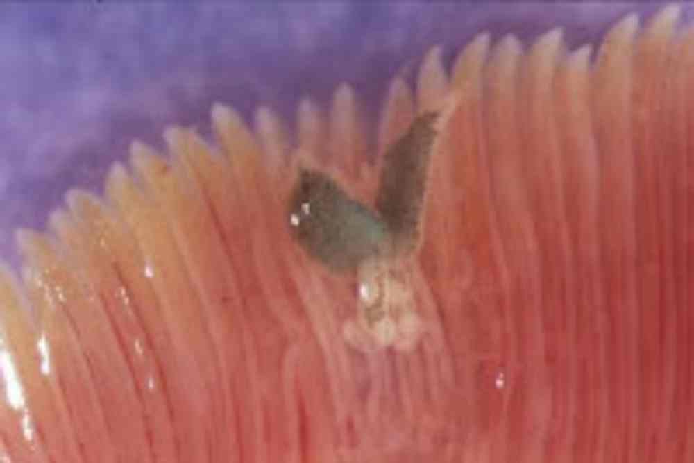
Diplozoon paradoxum is a unique and intriguing parasitic worm that inflicts gill and skin diseases in fish, known as Diplozooniasis.
This monogenean parasite has a distinctive X-shaped body with two heads and two hind parts, making it a fascinating specimen for study. Its life cycle is remarkable, involving a complex process of larval combination, resulting in “Siamese twin” parasites that cannot be separated.
Diplozoon paradoxum settles between the gill sheets of fish, hindering their breathing and causing excessive mucus production, leading to respiratory distress and erratic swimming behavior in heavily-infested fish.
In this article, we will explore the morphology, life cycle, symptoms, and treatment of Diplozoon paradoxum, shedding light on this unique parasitic worm and its impact on fish health.

Morphology of Diplozoon paradoxum
The Diplozoon, a unique and intriguing type of monogenean parasite, is like a dynamic duo in one body. With a visible size of 4-5 mm, it catches the eye with its X-shaped body sporting two heads and two hind parts.
Its body is bilaterally symmetrical and flattened from top to bottom, with an intestinal caecum present for digestion.
Unlike traditional suckers, it boasts a sucker-like structure at its anterior end, near its mouth opening, equipped with a sucking disc and an esophagus.
The Diplozoon’s has an attachment organ called opisthaptor situated at the posterior end of its body.
This remarkable structure is made even more efficient by the presence of toluidine blue, a Resilin-like substance found in the exoskeleton of arthropods.
It uses this opisthaptor to cling onto specific sites, primarily the gills of fish, where it becomes an ectoparasite causing gill and skin diseases known as “Diplozooniasis”.
It tends to prefer the dorsal part of the branchial arch, and the highest number of Diplozoons are found there.
In terms of reproduction, the Diplozoon is a hermaphrodite, possessing both male and female reproductive organs. It follows a direct life cycle, utilizing only one host for its survival.
Its unique body characteristics and fascinating attachment organ make the Diplozoon a truly remarkable parasite in the world of monogenean worms.
Life Cycle of Diplozoon paradoxum
The life cycle of Diplozoon paradoxum is a tale of unique simplicity, as it bypasses the need for changing hosts.
It all begins when the adult parasite produces eggs that are large, oval, and operculate, measuring 0.1-0.2 mm in size.
These eggs are then affixed to the gills of a fish by a long thread, and from them, larvae hatch and swim freely through the water in search of a suitable host.
Once the larvae find a fish, they mature and become sexually ripened, but not yet adult, youngsters known as diaspora.
These diaspora larvae are equipped with a sucking disc and a projection on opposite sides of their bodies, about halfway along their length.
In a fascinating twist, each larva inserts its projection into the sucker of another larva, and they grow into “Siamese twins” that are impossible to separate.
These united larvae, known as diporpa, are hermaphroditic, possessing both male and female reproductive organs. They mutually fertilize each other after combining, becoming one flesh in the most literal sense.
They are inseparable for the rest of their lives, as they cannot be separated once the union takes place.
At this stage, the diporpa have a size of 1.2 mm, and the process of combining to form a complete Diplozoon is an extraordinary feat that sets them apart from other parasites in the animal kingdom.
Geographical Distribution of Diplozoon paradoxum
The monogenean Diplozoon paradoxum is the parasite of various cyprinid fishes in Europe and Asia, Africa.
This monogenean had a progenitor from the Pacific. The progenitor of Octomacrum is the same but the distribution of Octomactum is limited in North America.
Biogeographic regions: Palearctic (native), oriental (native)
Habitat: Diplozoon paradoxum individuals parasitize cyprinid fishes. A study in Northern England found that it was absent entirely from fish of the genus Leuciscus.
In one area, the infection is present in some waters but absent in others and predominates in rivers rather than ponds or reservoirs.
D. paradoxum is randomly distributed on the gills, sides of the gill apparatus, hemibranches, and surfaces of primary lamellae of G. gobio, P. phoxinus, but prevails on median sectors of the gills of these hosts.
- Aquatic Biomes: Pelagic, benthic, lakes and ponds, rivers and streams, temporary pools
- Wetland: Marsh, swamp
- Other Habitat Features: Agricultural
Fish species affected by Diplozoon paradoxum
Diplozoon paradoxum has been found on the gills of several species of fish, such as Rui, Catla, Mrigal, Prussian carp, Minnows, Bream, Bitterling, Rudd, etc. In India, Diplozoon paradoxum has been reported from carp.
Causing Signs and Symptoms
When it comes to Diplozooniasis, the culprits behind the disease are none other than Diplozoon homoin or Diplozoon paradoxum, also known as the twin worm.
Among them, D. paradoxum takes up residence between the delicate gill sheets of its fish host, causing minimal harm.
However, its presence is not without consequences. Parts of the gill sheets where the flukes have settled become coated with a cloudy film, composed of slime and destroyed epithelial cells.
This triggers an excessive production of mucus in the gills, hindering the fish’s ability to breathe freely.
As a result, infected fish display signs of distress. They become lethargic, swimming sluggishly towards the water’s surface in an attempt to gulp air, a behavior known as “pipping”.
This desperate gasping for air at the surface is a telltale sign of severe respiratory distress, indicating the detrimental impact of the parasite on the fish’s ability to respire properly.
In cases where the infestation is severe, heavily-infested fish may exhibit erratic swimming behavior and become moribund, displaying a decline in vitality and strength.
The effects of Diplozoon paradoxum on fish are not to be taken lightly, as they can significantly impact the well-being and survival of the infected fish.
Diagnosis of Diplozoon paradoxum
Diagnosis of Diplozoon paradoxum typically involves several steps. Firstly, it usually requires a thorough examination of the affected fish, usually under a microscope. The following diagnostic methods may be used:
Macroscopic Examination: The adult Diplozoon paradoxum is a flatworm that is visible to the naked eye as a reddish or pinkish mass, resembling a “v”-shaped structure, attached to the gills or body surface of the fish.
This macroscopic examination can provide a preliminary diagnosis of Diplozoon paradoxum infection.
Microscopic Examination: A microscopic examination is necessary to confirm the diagnosis. A small piece of the reddish mass or gill tissue may be excised from the fish and placed on a microscope slide for examination under a compound microscope.
The microscopic examination typically involves staining the specimen with specific dyes, such as carmine or hematoxylin, to enhance the visibility of the internal structures of Diplozoon paradoxum.
Morphological Examination: The characteristic features of Diplozoon paradoxum include a flattened, symmetrical body with a “v”-shaped appearance, consisting of two large haptoral lobes at the anterior end, each with hooks for attachment to the host’s gill tissue, and a posterior part with reproductive organs.
These features can be examined in detail using a microscope to confirm the presence of Diplozoon paradoxum.
Molecular Diagnosis: In some cases, molecular methods may be employed to confirm the identification of Diplozoon paradoxum. DNA extraction from the parasite can be performed, followed by amplification of specific genetic markers through polymerase chain reaction (PCR) and sequencing to compare the obtained DNA sequences with known sequences of Diplozoon paradoxum.
Differential Diagnosis: It is important to rule out other similar parasites or pathologies that may cause similar symptoms or lesions in fish, such as other monogenean parasites, trematodes, or gill diseases caused by bacteria, fungi, or other parasites.
Prevention of Diplozoon paradoxum
When it comes to preventing Diplozooniasis, taking proactive measures is key. To keep your pond free from infection, consider stocking it with new fish, particularly fry or juveniles, after ensuring their disinfection.
One effective method is to drain the pond and treat the bottom with quicklime at a rate of 2 kg per hectare, allowing it to dry for 2-3 days. This treatment can effectively neutralize the parasite and its eggs, ensuring a clean and safe environment for the fish.
Alternatively, applying a disinfectant without drying the pond, at a recommended rate of 2.5 tons per hectare, can also be effective in preventing Diplozooniasis. This method ensures thorough coverage and eradication of the parasite from the pond.
Another preventive measure is to add methylene blue to the pond water at a rate of 1 gram per 10 cubic meters. Methylene blue has disinfectant properties and can help to control the spread of Diplozoon parasites in the pond.
By implementing these preventive measures, you can safeguard your fish and keep your pond free from Diplozooniasis, ensuring a healthy and thriving aquatic environment.
Diplozoon paradoxum Treatment
| Compound | Treatment method | Dose rate |
|---|---|---|
| Ammonium hydroxide (NH4OH) | Temporary bath (5-15 minutes) Short bath (2 minutes) | 500 ppm 1000 ppm |
| Sodium chloride(NaCl)(Common salt) | Dip (5 minutes) Bath (30 minutes) | 5% NaCl2% NaCl or 25-50 ppm |
| Potassium permanganate KMnO4 | Spray (part of the pond)Bath | 10 ppm 2 ppm or 3-5 ppm |
| Lysol | Dip (30 seconds) | 20 ppm |
| Copper sulfate (CuSO4.5H2O) | Bath or flush (30 minutes) | 100 ppm |
| Dipterex | – | 0.1-0.2 ppm |
| Malachite green | Spray (pond) | 0.15 ppm (should be continued for 3 days) |
References
- Wiles M. (1968). The occurrence of Diplozoon paradoxum Nordmann, 1832 (Trematoda: Mongenea) in certain waters of northern England and its distribution on the gills of certain Cyprinidae. Parasitology, 58(1), 61–70. https://doi.org/10.1017/s0031182000073418
- Wong, W. L., & Gorb, S. N. (2013). Attachment ability of a clamp-bearing fish parasite, Diplozoon paradoxum (Monogenea), on gills of the common bream, Abramis brama. The Journal of experimental biology, 216(Pt 16), 3008–3014 https://doi.org/10.1242/jeb.0
- Rolbiecki L. (2001). Topographic specificity of Diplozoon paradoxum nordmann, 1832 (Monogenea: Diplozoidae) in the bream, Abramis brama (Linnaeus, 1758) in the Vistula Lagoon, Poland. Wiadomosci parazytologiczne, 47(4), 687–691.
- Sebelov, S., Kuperman, B., & Gelnar, M. (2002). Abnormalities of the attachment clamps of representatives of the family Diplozoidae. Journal of helminthology, 76(3), 249–259. https://doi.org/10.1079/JOH2002133
- Wong, W. L., Michels, J., & Gorb, S. N. (2013). Resilin-like protein in the clamp sclerites of the gill monogenean Diplozoon paradoxum Nordmann, 1832. Parasitology, 140(1), 95–98. https://doi.org/10.1017/S0031182012001370
