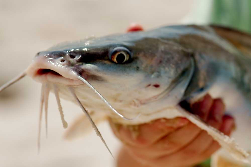
Viral diseases are an increasing problem for both wild and farmed fish populations globally.
One such viral disease is channel catfish viral disease (CCVD), which is caused by a member of the family Herpesviridae.
This disease can cause mortality in both juvenile and adult fish, and has been found in channel catfish in the United States, Canada, and Europe.
Channel Catfish Viral Disease (CCVD) is a serious threat to the channel catfish industry. It’s and acute hemorrhagic infection in channel and blue catfish.
The disease was first identified in 1980s, and has since spread to other states. The virus is believed to be spread through contact with contaminated water or food.
The disease is caused by a virus called channel catfish virus (CCV). Infected fish often display symptoms such as hemorrhage, exophthalmia, lethargy, appetite loss, and mortality.
CCVD is a serious threat to the channel catfish industry, and control measures are urgently needed.

Channel Catfish Virus
Channel catfish virus (CCVD) is a major virus with economic losses in countries where channel catfish (Ictalurus punctatus) are farmed.
Although this virus was isolated back in the 1980s, it took approximately five decades before the virus was studied using molecular tools, lagging far behind other pioneer herpesviruses.
Viruses are tiny infectious agents that can only replicate inside the cells of a living host. Channel catfish viral disease (CCVD) is a viral disease that affects channel catfish.
The disease is characterized by white lesions on the skin and fins, and internal organs such as the liver and spleen.
The RNA virus replication cycle proceeded between 10 and 33 degrees Celsius, but not higher. The highest growth was between 25 and 33 degrees Celsius.
Channel Catfish Viral Disease Symptoms & Signs
–Channel catfish herpesvirus infection resulted in the presence of intranuclear inclusions and extensive syncytium formation.
-Reduced feeding activity most likely the first sign and in high mortality for fry and juvenile with severe infection.
-The exophthalmos (popeye) is accompanied by hemorrhaging of the fins and ventral abdomen.
-Swelling of abdominal cavity contains fluid contaminants.
-Dark and enlarged spleen.
-Pale and enlarged kidney.
-Large numbers of fish aggregate around the edges of hatching troughs or pools, remaining motionless in a head-up, tail-down position.
Diagnosis of Channel Catfish Viral Disease
A microscopic examination of viruses by transmission electron microscopy revealed that they are about 100 nm in diameter in microbes.
The pathologist observed necrosis and hemorrhage in the kidney and liver tissue, the spleen and gastrointestinal tract, and hemorrhages in these areas.
Comparing a channel catfish ovary cell line to a brown bullhead cell line for their take on replication and detection of channel catfish virus (CCV) revealed that the cell line derived from the line was successful.
The Channel catfish virus PCR assay makes it possible to diagnose acute congenital virus. The method identifies the CCV DNA in tissues of acutely infected fish such as the spleen, stomach, intestine, blood, brain, and kidney.
Channel Catfish Viral Disease Treatment
The soluble antigen (envelope) from formidably channel catfish virus (CCV) was used for research as an processed into a vaccine for channel catfish viral infections since 1989.
Channel catfish immature eggs and newly hatched fry were vaccinated by immersion. A booster was administered to subgroups that were 2 weeks old after vaccination.
1 to 4 days old the eggs of channel catfish Ictalurus punctatus and 1 week old fry were vaccinated by immersion. A booster was given to subgroups of fry 2 weeks after vaccination.
References
- Kucuktas, H., & Brady, Y. J. (1999). Molecular biology of channel catfish virus. Aquaculture, 172(1-2), 147-161.
- Wolf, K., & Darlington, R. W. (1971). Channel catfish virus: a new herpesvirus of ictalurid fish. Journal of Virology, 8(4), 525-533.
- Bowser, P. R., & Plumb, J. A. (1980). Channel catfish virus: comparative replication and sensitivity of cell lines from channel catfish ovary and the brown bullhead. Journal of Wildlife Diseases, 16(3), 451-454.
- Plumb, J. A. (1971). Tissue distribution of channel catfish virus. Journal of Wildlife Diseases, 7(3), 213-216.
- Gray, W. L., Williams, R. J., & Griffin, B. R. (1999). Detection of channel catfish virus DNA in acutely infected channel catfish, Ictalurus punctatus (Rafinesque), using the polymerase chain reaction. Journal of Fish Diseases, 22(2), 111-116.
- Plumb, J. A., Gaines, J. L., Mora, E. C., & Bradley, G. G. (1974). Histophathology and electron microscopy of channel catfish virus in infected channel catfish, Ictalurus punctatus (Rafinesque). Journal of Fish Biology, 6(5), 661-664.
- Gray, W. L., Williams, R. J., Jordan, R. L., & Griffin, B. R. (1999). Detection of channel catfish virus DNA in latently infected catfish. Journal of General Virology, 80(7), 1817-1822.